Figure 1.
Fundus photograph of the left posterior pole shows the characteristic presentation of choroidal rupture. The involvement is temporal to the macula, does not encroach near the optic disc, and presents with pigmentary remodeling.
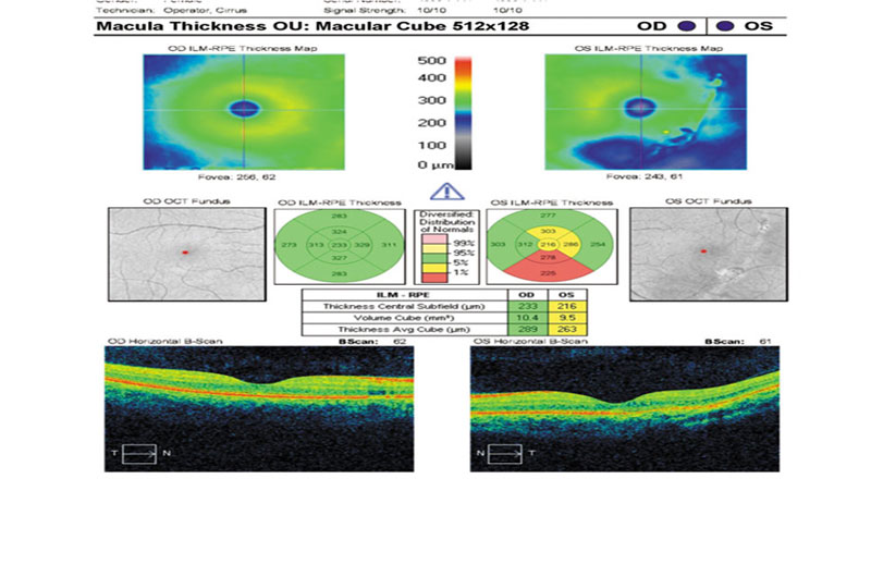
Figure 2. Macular optical coherence tomography (OCT) of each eye shows the contrast between
the involved area of the left eye compared with
that of the right eye. Note on the cross-sectional presentation running from nasal to temporal areas how the left eye shows general thinning of the retinal layers on the temporal aspect. Greater detail is present in other figures.

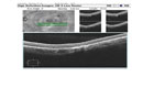
Figure 3. High-resolution scans through
the lesion demonstrate outer retinal layer disruption and retinal pigment epithelium (RPE)/ Bruch membrane remodeling consistent with choroidal rupture. No evidence for choroidal neovascularization was found. Careful evaluation also shows diminution of the inner retinal layers in the region immediately inner to the area of involvement. Note the discontinuity of the retinal nerve fiber layer (RNFL) and compare with the fundus appearance in Figure 1.
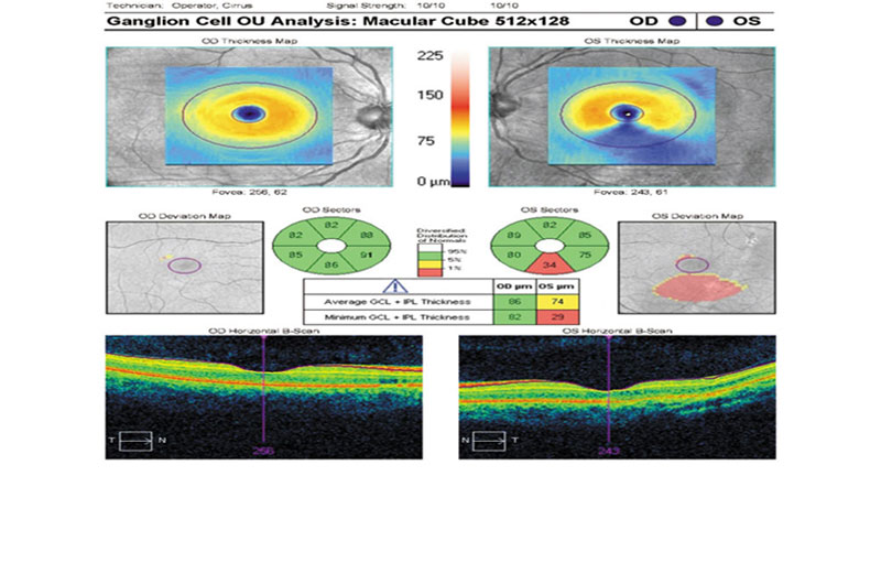
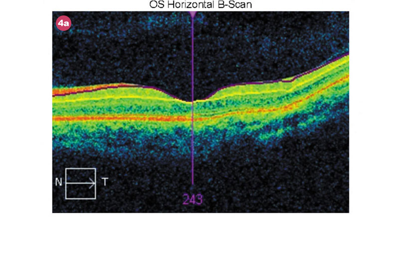
figure 4.
Reflection of the
inner retinal layer compromise is seen with the thinned ganglion-cell complex (GCC). This is due to loss of the RNFL alone. While there is perhaps the false impression of an almost absent GCC according to
the numbers, the cross-sectional images confirm regional attenuation of the RNFL. See the cross- sectional isolation in Figure 4a.